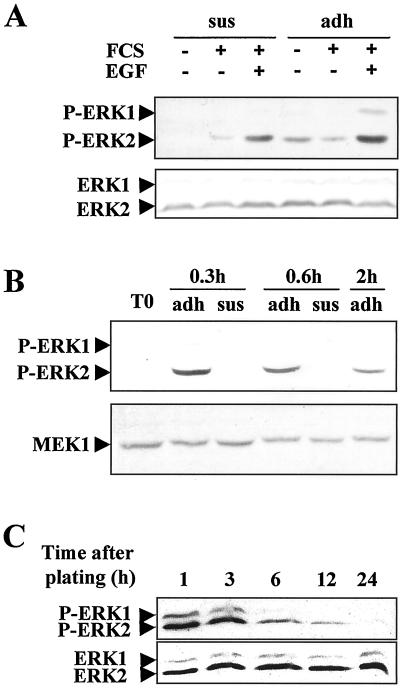
Figure 1.
Kinetics of ERK1/2 phosphorylation during early G1 in rat hepatocyte primary cultures analyzed by Western blotting using antibody directed against the phosphorylated forms. (A) Kinetics of ERK1/2 phosphorylation analyzed at the indicated times in suspended (sus) and adherent (adh) hepatocytes. Western blotting analysis was performed 3 h after hepatocyte isolation in suspended (sus) or adherent (adh) cells stimulated (+) or not (−) with FCS or EGF for 15 min. For detection of total ERK 1 and 2, a mixture of equal ratios of anti-ERK1 and anti-ERK2 antibodies was used. (B) Western blotting of ERK1/2 phosphorylation analyzed at the indicated times in hepatocytes seeded on a rigid film of type 1 collagen. T0, freshly isolated hepatocytes. The blot was stripped and reprobed with an anti-MEK1 antibody. (C) Kinetics of ERK1/2 phosphorylation in hepatocyte primary cultures continuously stimulated by FCS from seeding to 24 h. For the kinetics of total ERK1 and ERK2 proteins, a mixture of equal ratios of anti-ERK1 and anti-ERK2 antibodies was used. Experiments were performed at least three times.

