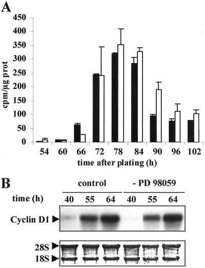
Figure 6.
DNA replication and cyclin D1 mRNA synthesis after PD98059 treatment. (A) Reversion experiment. Cultures were exposed for 42 h to the solvent control (black bar) or PD98059 (open bar). Then, the drug was removed and hepatocytes were stimulated by EGF. [methyl-3H]Thymidine incorporation into DNA was analyzed at the indicated times after growth factor stimulation. (B) Cyclin D1 mRNA was analyzed by Northern blotting in solvent control and PD98059-treated cells at the indicated times after drug removal. 18S and 28S rRNAs dyed by methylene blue coloration were used as the control. Experiments described were performed at least three times.

