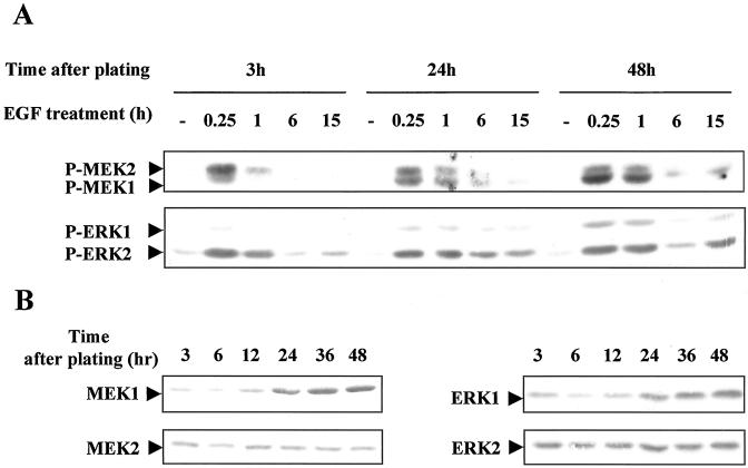
Figure 8.
Phosphorylation of different forms of MEK1/2 and ERK1/2 according to EGF stimulation in the G1 phase. (A) Hepatocytes were stimulated by EGF at different times in G1 phase progression (3, 24, and 48 h after plating). Phosphorylation of MEK1/2 and ERK1/2 was analyzed 0.25, 1, 6, and 15 h after the growth factor stimulation. −, control cells before stimulation at the indicated times. (B) Detection of total MEK1, MEK2, ERK1, and ERK2 proteins in hepatocytes during G1 progression analysis. Western blottings were performed using antibodies directed against each isoform. These data are representative of three different experiments.

