Figure 3.

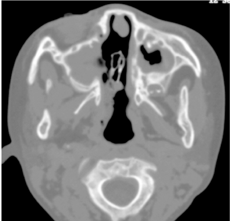
Fungal sinusitis of a four-year-old girl with myeloid leukaemia. CT of the sinuses revealed opacification of the maxillary sinuses with bone destruction of the medial wall. Mucor species was isolated from maxillary sinus washout.

Fungal sinusitis of a four-year-old girl with myeloid leukaemia. CT of the sinuses revealed opacification of the maxillary sinuses with bone destruction of the medial wall. Mucor species was isolated from maxillary sinus washout.