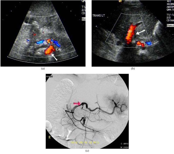
Figure 2.
Hepatic artery thrombosis after LDLT. (a) Colour Doppler US showed patent coeliac artery (arrow). (b) Absence of flow and Doppler signal seen at the expected location of the hepatic artery (arrow). PV: Portal vein. (c) Digital subtraction angiogram of the coeliac axis confirmed thrombosis of the hepatic artery (arrow).

