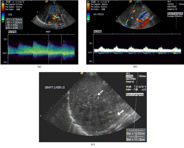
Figure 3.
Hepatic artery stenosis after liver transplantation with cholangitic abscess. (a) Colour Doppler US at post-stenotic segment of hepatic artery showed turbulent flow and elevated peak systolic velocity. (b) Tardus-parvus waveform at pre-stenotic segment of hepatic artery with dampened flow. (c) Hypoechoic cholangitic abscess at right lobe liver graft due to hepatic ischaemia (arrows).

