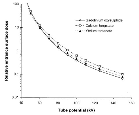
Figure 6.
Graphs of relative entrance surface dose against tube potential to obtain an image for a 200 mm thickness of soft tissue with three phosphors used in radiographic cassettes; gadolinium oxysulphide, calcium tungstate and yttrium tantanate. All calculations are performed for 200 μm thick phosphor layers and an X-ray beam filtered by 2.5 mm of aluminium.

