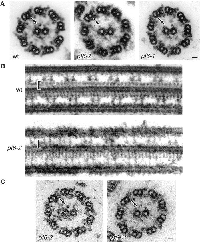
Figure 2.
Electron microscopic analysis of axonemes from wild-type (wt) and mutant Chlamydomonas cells. (A) Transverse sections of flagellar axonemes reveal that the C1-1a projection present in wild-type samples is missing from pf6-2 and pf6-1 preparations (arrows). (B) Longitudinal images reveal two rows of projections repeating at precise 16-nm intervals in wild-type samples, but one row of projections is missing in the pf6–2 axonemes (brackets). The observed central pair microtubule is identified as C1 based on the length of the remaining associated projections. (C) Rescue of the pf6 mutant motility defect by transformation with a clone containing a full-length wild-type copy of the PF6 gene is also accompanied by restoration of the C1-1a projection in both pf6-2 and pf6-1, as observed in axoneme cross-sections (arrows). Bars, 25 nm.

