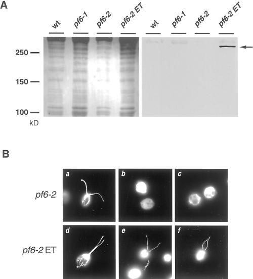
Figure 8.
Localization of the PF6 gene product. (A) Whole axonemes isolated from wild-type (wt), pf6-1, pf6-2, and a pf6-2 strain rescued with the epitope-tagged PF6 gene (pf6–2 ET) were separated on 6% acrylamide gels, blotted onto polyvinylidene difluoride, and stained with a reversible total protein stain (left) before immunolabeling with an antibody directed against the nine-amino acid HA epitope (right). The HA antibody recognized a single polypeptide present only in the pf6-2 ET sample that migrates slightly larger than 250 kDa in this gel system (arrow). (B) Indirect immunofluorescent localization of the PF6 gene product. pf6-2 (a–c) and pf6-2 ET (d–f) cells were labeled with antibodies against either α-tubulin (a and d) or the HA-epitope (b, e, and f). All of the recorded cells had flagella similar to those depicted in a and d. The tagged PF6 polypeptide is present along the entire length of the axoneme in pf6-2 ET cells (e and f). Control samples labeled with secondary antibody alone (c) demonstrate background cell body autofluorescence.

