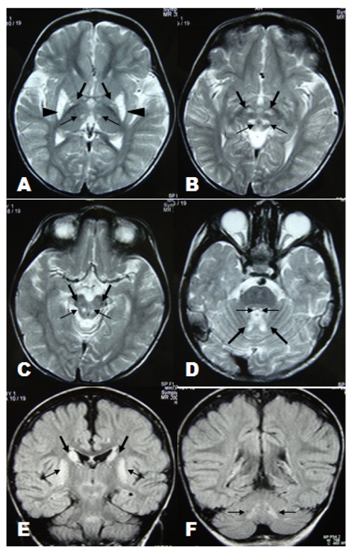
Figure 1.

(A): Axial T2-weighted image through the basal ganglia shows symmetrical hyperintense lesions involved thalamic posteromedial ventral nuclei (thin arrow), globus pallidi (thick arrow) and putamina (arrowhead). (B-D): Axial T2-weighted images through the brainstem show symmetrical involvement of reticular formation of midbrain (thin arrow in B), subthalamic nuclei (thick arrow in B), substantia nigra (thick arrow in C), dorsal midbrain (thin arrow in C) central tegmental tracts (thin arrow in D) and cerebellar nuclei region (thick arrow in D). (E-F): Coronal FLAIR images show symmetrical involvement of head of caudate nuclei (thick arrow in E), putamina (thin arrow in E) and dentate nuclei (thin arrow in F).
