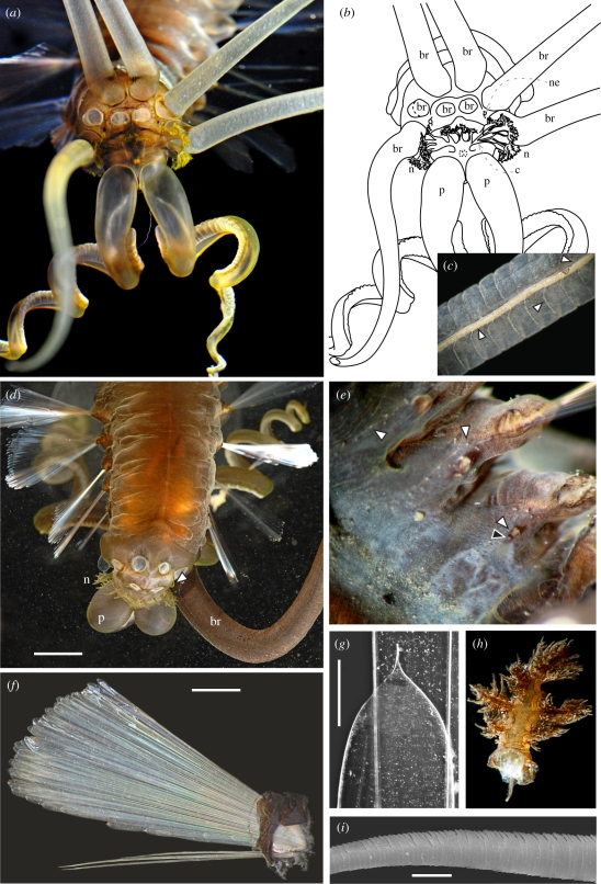
Figure 2.
Teuthidodrilus samae, gen. and sp. nov. (a and b) Anterior view of paratype 5 showing five attached branchiae (br), nephridiopores (black papillae, ne), branched nuchal organs (n) and the ciliary ridge joining them (c), and palps (p). (c) Shaft of elongate branchia from holotype showing longitudinal vessel (lower arrow), circular vessels (centre arrow) and nerve (upper arrow). (d) Dorsal view of holotype showing attached palps, branchial scars, branched nuchal organs, bubbled remains of the gelatinous sheath and nephridiopore (arrow). (e) Ventral view of paratype 3 showing notopodial (right) and neuropodial lobes (that of third chaetiger, lower white arrow), gonopore (black arrow), row of papillae (top right arrow) between noto- and neuropodial lobes and row of papillae (top left arrow) lateral across ventrum of segment. (f) Parapodium from holotype showing notochaetal fan (top) and neurochaetae (bottom). (g) Tip of notochaeta from holotype; 10× magnification is not sufficient to resolve the spinous nature of the distal tip. (h) Dissected free-standing, nuchal structure from paratype 4, frilly. (i) Neurochaeta distal shaft from holotype seen with scanning electron microscopy. Scale bars, d = 4 mm, f = 3 mm, g = 0.2 mm and i = 10 µm. (Online version in colour.)

