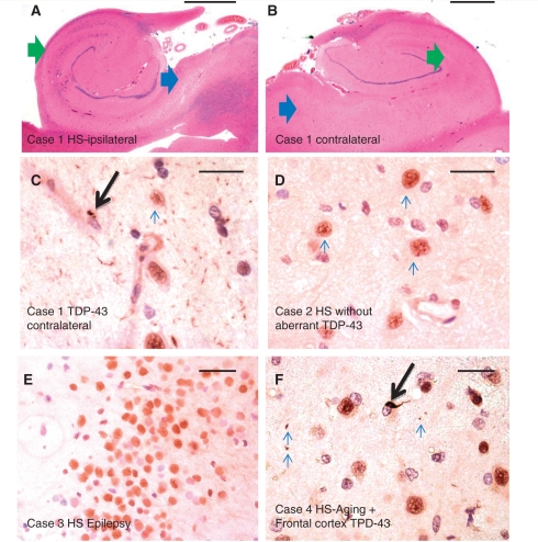
Figure 2.
Features of HS-Ageing and TDP-43 immunohistochemistry. A 97-year-old female (Case 1) had hippocampal features that, with haematoxylin and eosin staining, met criteria for hippocampal sclerosis on the right (A) but not the left (B) side. Green arrows show CA1, blue arrows show subiculum. Immunohistochemistry for TDP-43 showed clear evidence of aberrant TDP-43 on the left side (C), confirming that the process was bilateral. Case 2 (D) is an 88-year-old male with bilateral hippocampal sclerosis pathology but not Alzheimer’s disease or cerebrovascular disease, without aberrant TDP-43 staining. Shown here is the normal pattern of TDP-43 staining in neurons in the CA1 field of the hippocampus. Case 3 (E) is from a 34-year-old female with history of seizures and right mesial temporal sclerosis. Shown is a portion of the dentate granule cells, which like the rest of the hippocampectomy specimen (and all the other hippocampal sclerosis–seizures cases) showed no evidence of aberrant TDP-43 in cytoplasm or neurites. Case 4 (F) is from a 97-year-old APOE 3/3 male with Alzheimer’s disease and bilateral hippocampal sclerosis. Shown is a representative high-power field from frontal lobe (Brodmann area 9), with sparse aberrant TDP-43 immunohistochemistry including an intraneuronal inclusion (arrow) and a few scattered neurites (smaller arrows). Four of 14 stained frontal cortices from cases with HS-Ageing–TDP also showed scattered immunopositivity in frontal cortex. Scale bars = 1 mm (A, B and F), 50 microns (C–E). HS = hippocampal sclerosis.

