Fig. 2.
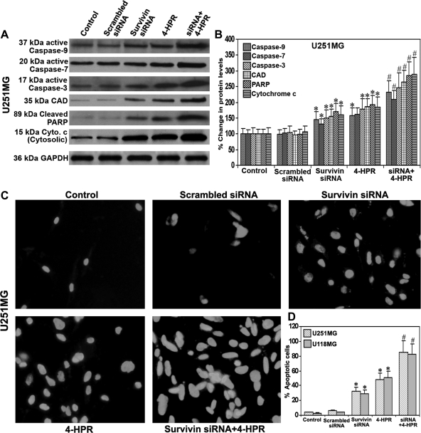
Activation of caspases and induction of apoptosis in glioblastoma cells. Treatments (48 hours): control, scrambled siRNA, survivin siRNA, 1 µM 4-HPR, and survivin siRNA + 1 µM 4-HPR. (A) Representative Western blots for active subunits of caspase-9, caspase-7, caspase-3, CAD, cleaved PARP, and cytosolic cytochrome c in the cell lysates of U251MG cells. Cleaved PARP was determined in the nuclear fraction. Mitochondria were isolated from the total cell lysate, and cytochrome c levels were determined in the cytosolic fraction. Western blots were reprobed for GAPDH to demonstrate that equal amounts of proteins were loaded in all lanes. (B) Quantitative evaluation of percent changes in the protein levels. Western blot images were quantified using Gel-Pro analyzer software. Data are mean ± SD of 6 independent experiments (*P < .001 compared with the mean values of control and #P < .001 compared with the mean values of survivin siRNA- or 4-HPR-treated samples). (C) TUNEL staining for the detection of apoptotic cells. (D) Quantitation of TUNEL positive cells using Image-Pro Discovery software. Data are mean ± SD of 6 independent experiments in each group (*P < .001 compared with the control mean values and #P < .001 compared with the survivin siRNA or 4-HPR mean values).

