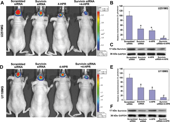
Fig. 6.
Inhibition of intracerebral tumorigenesis in nude mice after the treatments. (A and D) In vivo imaging of intracerebral tumorigenesis in nude mice. U251MG or U118MG cells (1 × 10⁶ cells) stably transfected with luciferase gene were injected into the intracerebrum of nude mice. Beginning from day 3, the mice were injected either with survivin siRNA plasmid vector (5 µg DNA/injection/mouse), 4-HPR (1 µg/injection/mouse), or both agents together for 20 days on alternate days at the site of tumor cell implantation. Then, the mice were injected intraperitoneally with 100 µL (50 mg/mL) of luciferin and visualized for the effect of treatments using Xenogen IVIS-200 imaging system. The data are representative of 6 sets of mice in each group. The background bioluminescence signal from an untreated mouse is about 1.5e + 05 photons on Xenogen IVIS-200 imaging machine. (B and E) Quantitative presentation of relative bioluminescence as percentage of photons in both U251MG and U118MG tumors. Data are mean ± SD of 6 animals in each group (*P < .001 compared with the scrambled siRNA mean values and #P < .001 compared with the survivin siRNA or 4-HPR mean values). (C and F) Western blots for survivin protein levels in the brain tumor tissues after the treatments. The blots were reprobed for GAPDH content to demonstrate that same amount of protein was loaded in each lane. The data are representative of 4 independent experiments.

