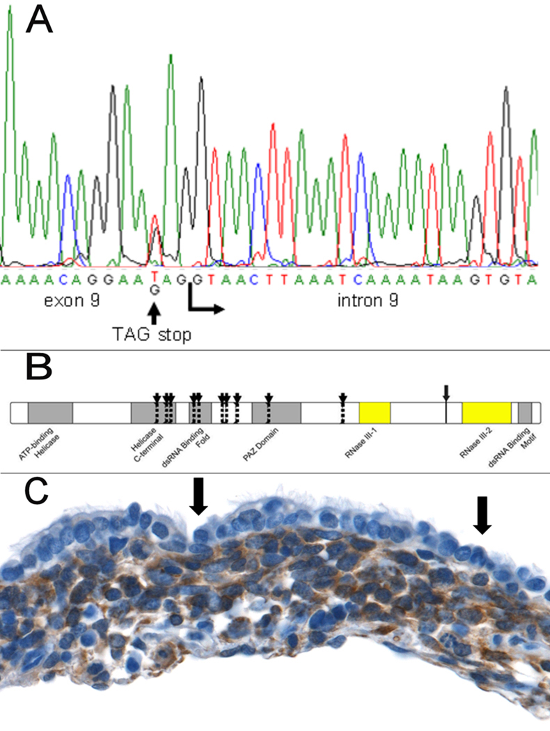
Figure 1. DICER1 mutations and expression in PPB.
(A) DICER1 sequence alteration identified in one of the families in the linkage study. (B) Protein locations of DICER1 mutations in 11 PPB families. Vertical dotted lines with arrows indicate the location of truncating or insertions/deletions in 10 families. Each of these occurs proximal to the RNase III domains. The larger arrow with the solid line marks the location of the missense mutation between the RNase III domains. (C) DICER1 protein is absent in benign-appearing tumor-associated epithelium (arrows) and is present in the mesenchymal tumor cells forming the cambium layer underneath (anti-DICER1 with brown chromagen and hematoxylin counterstain; original magnification x400).

