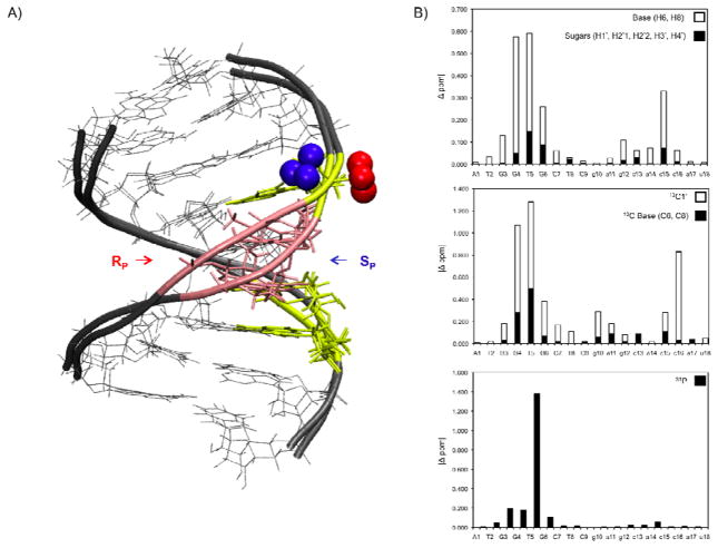
Figure 8.
A) Overlay of RP and SP hybrid structures, color coded according to chemical shift differences between the two modified hybrids. RP and SP hybrids aligned by all heavy atoms (except boron). Nucleotides (Σ (|H1′| |H2′1| |H2′2| |H3′| |H4′| |H6/H8| |13C1′| |13C6/13C8 |)
 > 1.00 ppm, 1.00 ppm <
 > 0.50 ppm,
 < 0.50 ppm. B) RP and SP hybrid chemical shift difference plotted by residue.
1H (top) closed box denotes Σ (|H1′| |H2′1| |H2′2| |H3′| |H4′|), 13C (middle), 31P (bottom) all Δ ppm. T5–P–G6 (31P) display the largest difference in chemical shift between the two modified hybrids (Δ 1.2 ppm).

