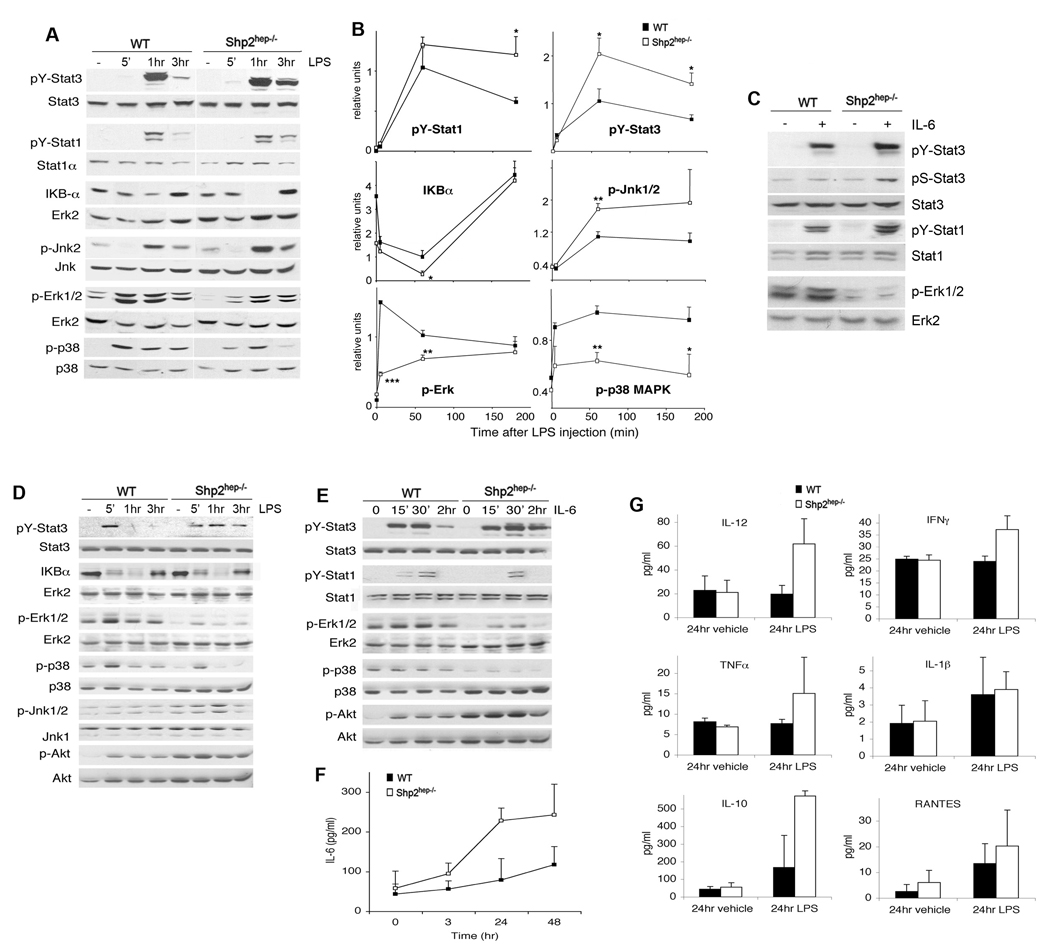
Figure 3. Altered hepatic LPS and IL-6 signaling events in vivo and in vitro.
(A) Mice were injected with LPS, and pY-Stat1, pY-Stat3, p-Jnk1/2, p-Erk1/2, and p-p38 MAPK were assessed by immnoblotting with their protein levels as controls. IκBα degradation was assessed, using Erk2 protein as control.
(B) The phospho-signals were quantified and normalized against total liver protein amounts. IκBα protein amounts were normalized against Erk2 The relative signal levels were determined by setting the value of the control at 1 hr as 1 unit. (n = 3–6, * p<0.05, ** p<0.01 for WT versus Shp2hep−/−).
(C) Immunoblotting of liver lysates was performed 5 min after injection of IL-6 (5 µg) into portal vein. pY-Stat3 and pS-Stat3, and p-Erk levels were assessed.
(D, E) pY-Stat3 and pS-Stat3, p-Jnk1/2, p-Akt (Ser473), p-Erk, p-and p38 MAPK were assessed by immunoblot analysis with protein levels as control. The IκBα degradation was compared to Erk2 protein level.
(F–G) Amounts of inflammatory cytokines were quantified in supernatants from WT and Shp2hep−/− hepatocytes (n = 3).

