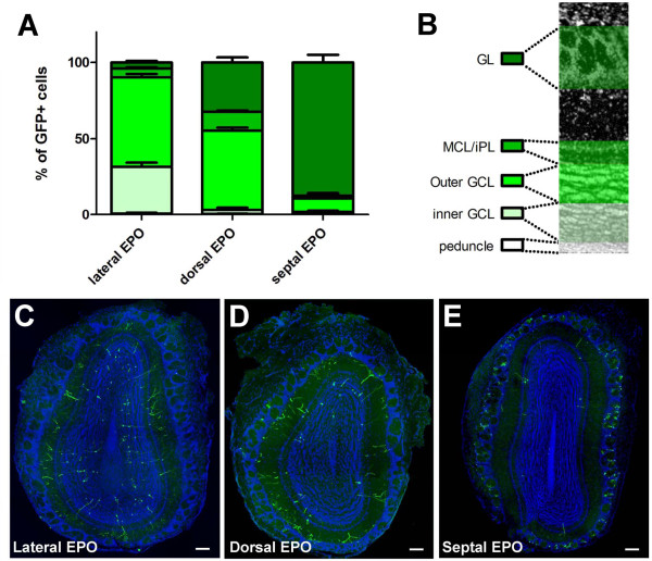
Figure 4.
Radial glial cells located in defined walls of the postnatal lateral ventricle produce neurons that migrate to distinct layers of the olfactory bulb. (A,B) Quantification of the percentage of GFP+ newborn neurons in defined layers of the OB at 21 dpe. The OB layers are illustrated in (B). Error bars represent standard error of the mean. (C-E) Representative overviews of the distribution of newborn neurons in the OB of laterally, dorsally, and septally electroporated animals. DAPI (blue) was used as a nuclear counterstain. Scale bars: 100 μm. GCL, granule cell layer; EPO, electroporation; GL, glomerular layer; iPL, internal plexiform layer; MCL, mitral cell layer.

