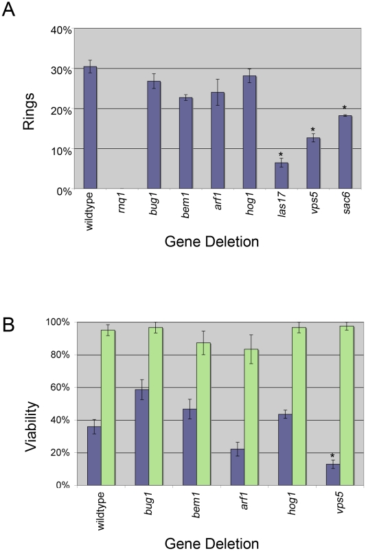
Figure 3. Ring formation and viability.
A. [PIN +] cells containing Sup35PD-GFP were induced in copper containing liquid media for 24 hours. The percentage of cells containing rings was determined by counting more than 300 cells from at least one transformant from each independent knockout line (see Table S3) for a total of three transformants per deletion. Bars indicate standard error and (*) indicates deletion strains that showed a significant difference in the percentage of cells containing rings compared to wildtype (p<0.01 unpaired t-test). B. Ring containing cells (purple bars) or cells with diffuse cytoplasmic fluorescence (green bars) were isolated by micromanipulation, placed on rich media and assayed for growth. At least 20 ring containing cells and 10 diffuse cells, from at least one transformant from each independent knock out line for a total of three transformants, were tested. Bars indicate standard error and (*) indicates the deletion strains that showed a significant difference in viability compared to wildtype strains (p<0.01 unpaired t-test).

