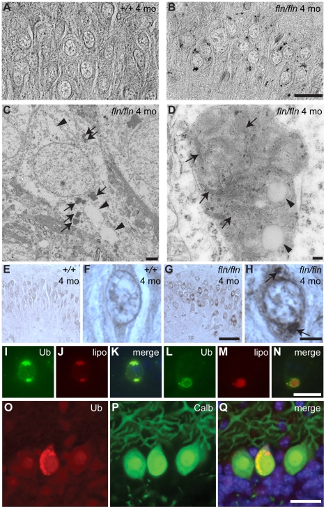
Figure 5. Lipofuscin and ubquitylated protein accumulation in CerS1-deficient neurons.
(A–B) Sections of cerebella from four-month-old wild type (+/+, A) and fln/fln (B) mice were stained with Luxol Fast Blue. Images are converted to gray-scale for clear visualization of punctate staining. Images of the hippocampal CA3 region are shown. (C–D) Electron micrographs showing electron-dense deposits in fln/fln neurons at five months of age. An overview of a hippocampal neuron (C) showing vacuoles (arrowheads) and electron-dense deposits (arrows), and a lipofuscin structure from a cortical neuron (D) showing internal membrane structures (arrows) and vacuoles (arrowheads). (E–H) Ubiquitin immunohistochemistry of the hippocampal CA3 region from 4-month-old wild type (+/+, E, F) and fln/fln (G, H) mice. Details are shown in higher magnification (F, H). Conspicuous ubiquitylated accumulations in an fln/fln neuron are marked with arrows (H). (I–N) Ubiquitylated proteins (Ub; I, L) and autofluorescent lipofuscin (lipo; J, M) in a hippocampal neuron (I–K) and a cortical neuron (L–N) of a four-month-old fln/fln mouse. Merged images (K, N) show the relative locations of the ubiquitin immunofluorescent stain and autofluorescence. (O–Q) Ubiquitin-positive inclusions in Purkinje cells of three-week old fln/fln mice. Sections were incubated with an antibody against ubiquitin (O) and calbindin D-28 (P). Merged images are shown (Q), with Hoescht dye counterstaining. Scale bars: 20 µm (A–B); 2 µm (C); 100 nm (D); 50 µm (E, G); 20 µm (F, H, I–N); 25 µm (O–Q).

