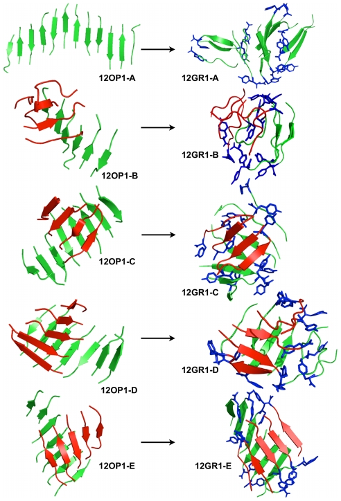
Figure 5. Structures obtained for the dodecameric simulations.
We show, on the left-hand side panel, representative structures obtained from the OPEP simulations and, on the right-hand side panel, representative structures obtained after all-atom MD refinements. 12OP1-A,-B,-C,-D and –E were extracted respectively at 222.5 K, 235.7 K, 250.8 K, 266.7 K and 283.4 K. 12OP1-A (top left structure) is a long flat beta-sheet. 12OP1-B to -E (second left to bottom left structures) are made of 2 beta-sheets facing each other. Monomers forming β-sheets in the initial state are colored red or green. These colors are kept in the final structure. The tyrosines are shown in blue sticks for the all-atom structures. During the all-atom MD simulation the structures tend to be more globular but the strands see no exchange between the β-sheets, i.e. the red and green β-sheets do not dissociate for the 12-mer system.

