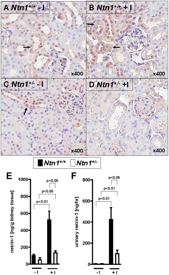
Figure 2. Immunohistochemical localization of renal netrin-1 and netrin-1 tissue content and urine concentration following ischemia in vivo.
Immunohistolochemical staining for netrin-1 in kidneys of Ntn1+/− mice or their respective age-, weight-, and gender-matched littermate controls (Ntn1+/+) following 30 minutes ischemia and 2 hours reperfusion. (A) Netrin-1 protein is mainly expressed in tubule cells of Ntn1+/+ mice under basal conditions without ischemia (−I) and (B) is increased following ischemia (+I). (C,D) This increase of netrin-1 expression following ischemia is attenuated in Ntn1+/− mice. Arrows indicate tubules with netrin-1 expression. (magnification 400×). (E) Renal and (F) urine netrin-1 content were assessed by ELISA (mean ± SD; n = 6–8).

