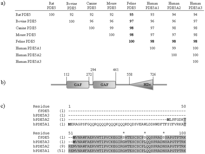
Figure 1. Sequence and Structural features of feline PDE5.
(a) Similarity between feline PDE5 and PDE5s from other species. The feline PDE5 sequence was obtain from the translation of the cloned cDNA sequence. Sequences of PDE5 from other species were obtained from GenBank. Multi-sequence alignments were calculated using Clustal W. The % identities between feline and other species are in bold. (b) Domain structure of the feline PDE5 protein. GAF indicates cGMP binding domain, and HDc the phosphohydrolase catalytic domain. Structure prediction was performed using the SMART software. (c) Alignment of protein sequences from the N-terminal region of feline and human PDE5 variants. The shaded area indicates identical residues and the residues in feline PDE5 that are different from those of human variants are noted with asterisks. The start codons are in bold faces and amino acid residue numbers are indicated.

