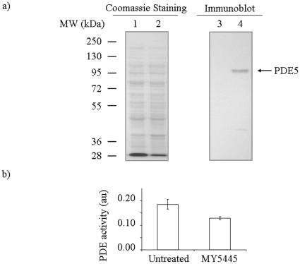
Figure 2. Immunoblotting and enzyme activity measurement of feline PDE5 expressed in E. coli.
(a) Immunoblot analysis and coomassie blue staining of whole cell extracts made from E. coli cells carrying feline PDE5 expression plasmid pT7-PDE5 or a control vector pSNAP-tag(T7). Lanes 1 and 3 are extracts from cells carrying pSNAP-tag(T7). Lanes 2 and 4 are extracts from cells carrying pT7-PDE5. Lanes 1 and 2 are Coomassie blue staining of the gel. Lanes 3 and 4 are immunoblot using an anti-human PDE5 antibody. (b) Measurement of PDE5 activity. PDE activity of the extracts from cells carrying pT7-PDE5 in the presence or absence of a PDE5 inhibitor, MY5445, was shown (see Method for details). Basal level PDE activity determined with extract from cells carrying pSNAP-tag(T7) was subtracted. The error bars indicate standard deviation (n = 4).

