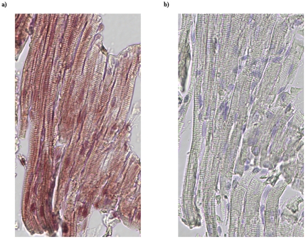
Figure 3. Detection of PDE5 expression in myocytes of feline RV by IHC staining.
Consecutive paraffin embedded RV tissue sections from control feline hearts were used to examine PDE5 expression. The section in a) was incubated first with a rabbit polyclonal anti-PDE5 antibody followed by HRP conjugated anti-rabbit secondary antibody and then developed using ImmPACT NovaRED as a substrate to visualize the bonded antibodies. Section b) was processed in parallel with a) but omitted the primary anti-PDE5 antibody. Hematoxylin QS was used as nuclear counterstaining.

