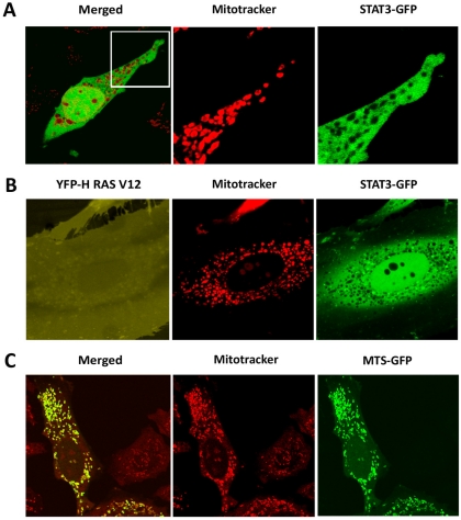
Figure 5. STAT3-GFP is excluded from mitochondria.
A) HeLa cells expressing STAT3-GFP were stained with MitoTracker Orange and the localization of STAT3-GFP and mitochondria was captured with live cell imaging. B) HeLa cells co-expressing STAT3-GFP and YFP-RasV12 were stained with MitoTracker Orange and live cell imaging identified the localization of YFP-RasV12, STAT3-GFP, and mitochondria. C) HeLa cells expressing MLS-GFP were stained with MitoTracker Orange and live cell imaging captured the localization of MLS-GFP and mitochondria. Images captured with Zeiss LSM 5 using maximal vertical resolution (<1 µm) and either 40× oil objective or 63× C-Apochromat (water) objective.

