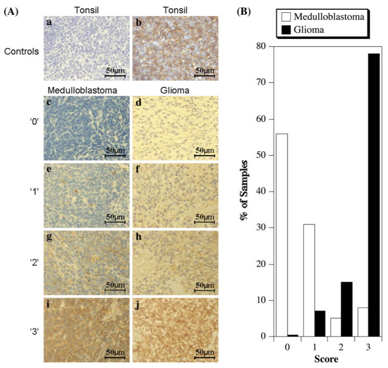
Fig. 2.

Immunohistochemistry analysis of medulloblastoma and adult glioma for HLA class I. a Representative sections showing staining with the heavy chain specific monoclonal antibody HC-10 (×40 magnification). Control tonsil tissue sections processed identically except for the absence (a) or presence (b) of primary HC-10 monoclonal antibody. Examples of medulloblastoma (c, e, g, i) or glioblastoma (d, f, h, j) sections graded as ‘0’ (c, d), ‘1’ (e, f), ‘2’ (g, h) and ‘3’ (i, j). b Relative distribution of HC-10 staining intensities in medulloblastoma and adult glioblastoma microarrays
