
Fig. 5.
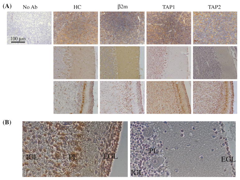
HLA class I expression in developing cerebellum. a Tissues from adult tonsil (top row), perinatal/early childhood (40 weeks of gestation through 3 years after the birth- middle row) or prenatal cerebellum were stained with heavy chain (HC)-, β2m, TAP1- and TAP2-specific antibodies and viewed at 20× magnification. For the perinatal/early childhood group, examples of negative staining were deliberately chosen, even though not all antibodies stained negative. b Higher magnification (×40) view of the prenatal (35 weeks of gestation; left) and 1 year old cerebellum specimens stained with HC-10 monoclonal antibody