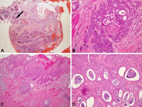
Fig. 1.
Histologic features of adenosquamous carcinoma. a Biopsy specimen showing a mixture of mature, keratinizing-type SCC and adenocarcinoma characterized by cells with higher nuclear to cytoplasmic rations and scattered gland formation (arrow) with basophilic mucin (100× magnification). b Punched out, rounded glandular spaces devoid of mucin (200× magnification). c Surface, keratinizing-type squamous cell carcinoma in situ and submucosal nests of cells with gland formation (100× magnification). d Florid gland formation with round, smooth edges and basophilic intraluminal and also intracytoplasmic mucin (200× magnification)

