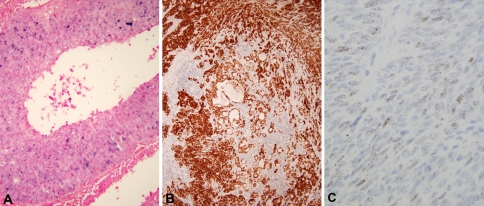
Fig. 2.
Immunohistochemical and in situ hybridization studies of adenosquamous carcinoma. a Positive DNA in situ hybridization showing granular, blue nuclear staining of tumor cells (200× magnification). b Positive p16 immunohistochemistry showing strong, diffuse nuclear and cytoplasmic staining of both squamous and glandular components of the tumor (100× magnification). c Positive RNA in situ hybridization showing granular, brown nuclear and cytoplasmic staining (400× magnification)

