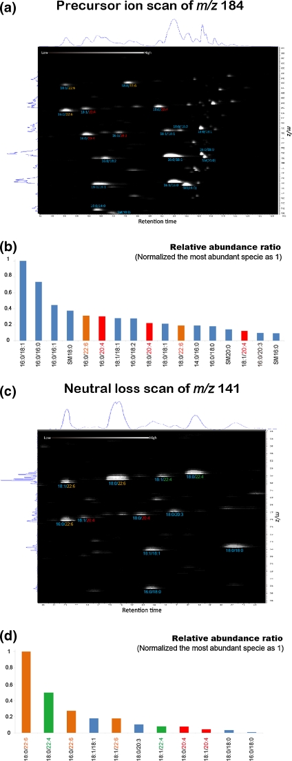
Fig. 2.
Identification of individual molecular species of focused phospholipid classes by precursor ion and neutral loss scanning of their head groups in the positive ion mode. The extracted total lipid mixture from the control postmortem human brain was subjected to precursor ion scanning of m/z 184 for detection of PCs and SMs (a, b) and neutral loss scan of m/z 141 for detection of diacyl form PEs (c, d). The 2D map (a, c) has the m/z value along the vertical axis and the retention time along the horizontal axis. The total ion chromatogram and averaged mass spectra were shown on the upper and left sides of the 2D map, respectively. From the 2D map, the relative ion counts of each PC, SM, and PE molecule species were calculated (The most intense species is shown as intensity at 1.) Detected PUFAs, namely, arachidonic acid, DHA, and docosatetraenoic acid, are shown as red, orange, and green characters, respectively

