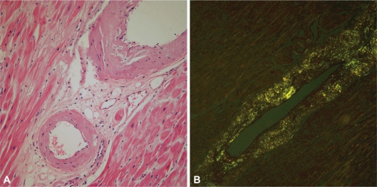
Fig. 3.
Light microscopic findings of vascular amyloid deposition. A: hematoxylin-eosin staining shows deposition of pale-staining amorphous material in both blood vessel and interstitium (original magnification ×100). B: congo red staining with cross-polarized microscopy shows that a green birefringence characteristic of amyloid is apparent (original magnification ×100).

