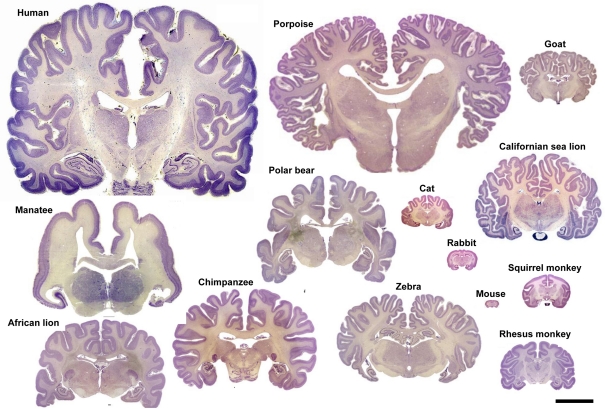
Figure 10.
Variation in brain size and patterns of convolutions. Coronal sections (25–40 μm thick; thionin staining) of the brain of several mammalian species. Adapted with permission from http://www.brains.rad.msu.edu, supported by the US National Science Foundation. Scale bar: 10 cm.

