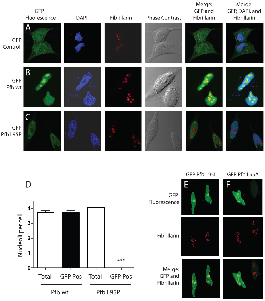
Figure 4. The L95P parafibromin mutation blocks nucleolar but not nuclear localization.
Hela cells were cultured in chamber slides and transfected with GFP vector control (A) or else GFP fusions with wild-type (Pfb wt) (B) or L95P mutant parafibromin (Pfb L95P) (C). Cells were then treated with the DAPI nuclear stain, immunostained using anti-fibrillarin antibody as a nucleolar marker, and analyzed by confocal laser fluorescence microscopy. Phase contrast and overlay (merge) of GFP fluorescent signal with the nucleolar marker without or with the nuclear stain are also shown. D. Quantitation of nucleoli per Hela cell transfected with GFP fusions with wild-type (Pfb wt) or L95 mutant parafibromin (Pfb L95P): total nucleoli counted if red on fibrillarin-only stained images; GFP-positive nucleoli counted if yellow on merging of GFP fluorescence and fibrillarin stained images. Means shown from pooling of 2 or 3 experiments per transfection, with 19 to 24 cells (nuclei) analyzed per experiment. ***, two-tailed P value <0.0001 compared to total, employing Student’s unpaired t test. E., F. Confocal laser fluorescence microscopic images of Hela cells transfected with GFP fusions with L95I mutant (Pfb L95I) (E) or L95A mutant parafibromin (Pfb L95A) (F), and analyzed as indicated, in A–C.

