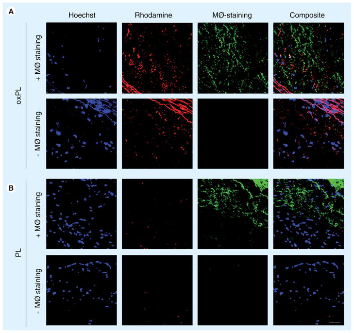
Figure 5. Confocal microscopy of atherosclerotic plaque.
Tissue samples were stained with cy-5 RAM 11 antibody (green pseudocolor) directed to macrophages. In addition, macrophage staining was omitted in order to confirm that there is no overlap between the two fluorescent signals. Vesicles were detected by red rhodamine fluorescence only in oxPL-injected animals. Hoechst staining was used to visualize nuclei (blue on merged images). Merged images showed colocalization of oxPL in macrophage-rich plaque areas, appearing in yellow composite. Bar represents 50 μm.
MØ: Macrophage; oxPL: Cholesteryl-9-carboxynonanoate phospholipid vesicles; PL: Phospholipid.

