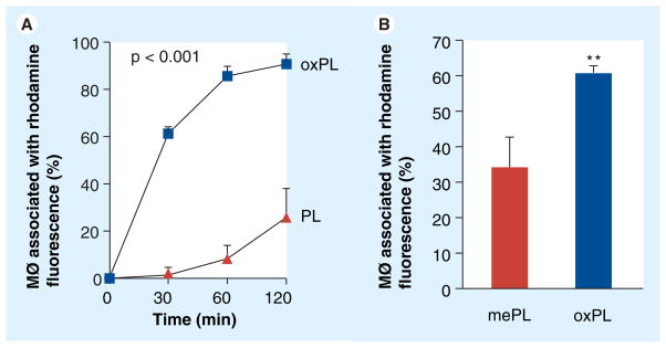
Figure 6. In vitro study of uptake of oxPL by human macrophages (MØ) as compared with phospholipid.
Binding of rhodamine-containing vesicles to macrophages was assessed by flow cytometry and presented as percentage of cells associated with rhodamine fluorescence. (A) Time course of vesicle uptake by macrophages. (B) Binding of oxPL as compared with control vesicles containing methyl ester-modified 9-CCN.
MØ: Macrophage; oxPL: Cholesteryl-9-carboxynonanoate phospholipid vesicles; PL: Phospholipid.

