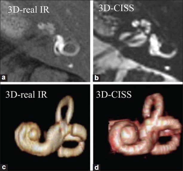
Figure 3.

MRI protocols in a patient with Meniere’s disease. High spatial resolution 3D-real inversion recovery (IR) image (a, 0.8 mm thick) and 3D-constructive interference in the steady-state (CISS) image (b, 0.4 mm thick), and their volume-rendered (VR) images (c, d). By comparing the perilymphatic VR image (c) and total lymphatic VR image (d), we can appreciate the degree of endolymphatic hydrops three dimensionally (Naganawa S, Nakashima T. Cutting edge of inner ear MRI. Acta Otolaryngol 2009;129:15-21)
