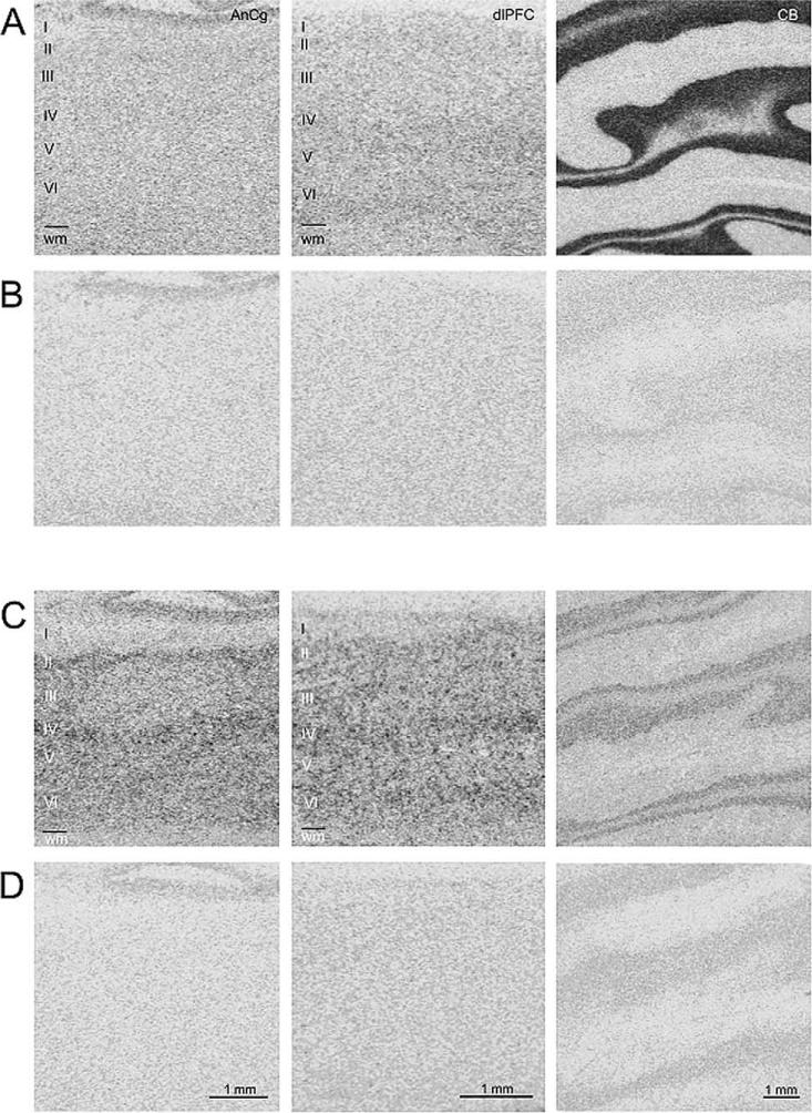
Fig. 3.
In situ hybridization histochemistry of PAK-3 and CHES1. The specific signals from 35S-labeled riboprobes relative to RNase-negative controls are shown for representative sections from AnCg, DLPFC, and CB. Rows A and C show specific signal for CHES1 and PAK-3, respectively, across labeled sections. Rows B and D show signal from RNase controls for CHES1 and PAK-3, respectively, of sections adjacent to those immediately above.

