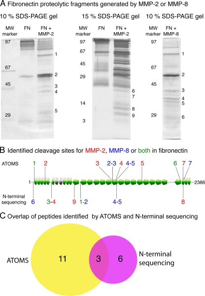
Fig. 4.
Comparative analysis of cleavage sites identified by ATOMS and N-terminal sequencing. Fibronectin-1 was cleaved by MMP2 or MMP8 after which the reaction mixtures were separated in two aliquots and subjected to ATOMS or SDS-PAGE and N-terminal sequencing by Edman degradation. A, SDS-PAGE gels of fibronectin-1 (FN) digested with MMP2 and MMP8. The band numbers are associated with the peptides sequenced and presented in Table II. Molecular weight markers are indicated on the left. B, schematic of the cleavage sites identified by ATOMS and N-terminal sequencing. Cleavage sites identified by ATOMS and N-terminal sequencing are labeled on top and bottom of the schematic, respectively. MMP2 cleavage sites are blue, MMP8 sites are red, and cleavage sites common to MMP2 and MMP8 are green. A Venn diagram of the number of cleavage sites identified by ATOMS (yellow) and N-terminal sequencing (magenta) is shown.

