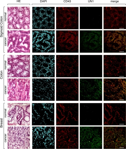
Fig. 9.
UN1-type CD43 glycoforms are expressed in cancer tissues. Serial cryosections of surgical specimens were derived from normal and cancer tissues of sigmoid colon, colon, and breast. For optical microscopy, sections were stained with hematoxylin and eosin (HE). For confocal microscopy, sections were stained with DAPI (blue), or the immunofluorescent antibodies CD43 (C-20) (red) and UN1 (green). Image superposition of UN1 and CD43 (merge) indicates colocalization by yellow color. Bars, 47.61 μm.

