Figure 2.
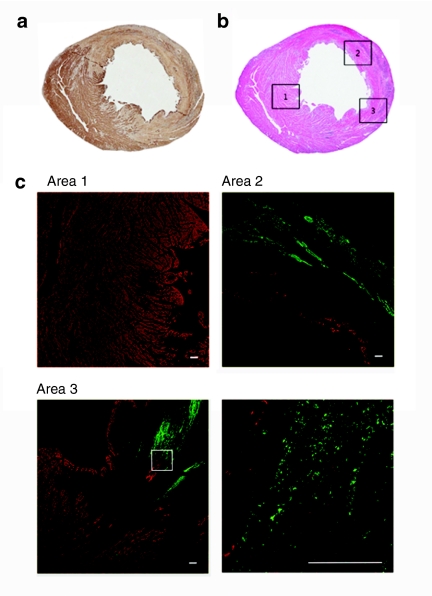
Microscopic features of bacterial tropism for infarcted myocardium. ΔppGpp S. typhimurium (2 × 10⁸ CFUs) was injected through the tail-vein into Sprague–Dawley rats with MI (n = 5, each group). (a) Distinct delineation between healthy myocardium and infarcted myocardium from heart after immunohistochemical detection of desmin at 5 days post inoculation. Positive detection of desmin corresponds to the healthy region (brown), while negative detection corresponds to an infarcted lesion. (b) Hematoxylin-eosin (H&E) staining of a cardiac cross-section that corresponds to desmin immunoreactivity. (c) Immunofluorescence staining of indicated H&E stained areas. Sections were stained with antidesmin antibody (red) and anti-Salmonella antibody (green). Bottom right panel is a magnification of the boxed area in the bottom left panel (area 3). Bars = 100 µm. CFU, colony forming units; MI, myocardial infarction.

