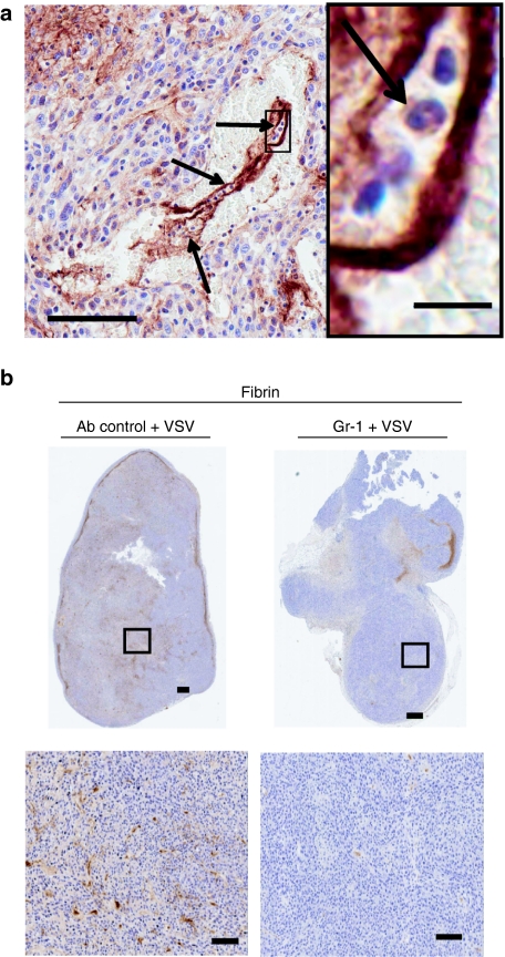
Figure 6.
Decreased clot formation is observed in tumors from neutrophil-depleted mice. (a) A fibrin clot within a tumor vessel. Arrows indicate neutrophils. Bar = 100 µm, magnification: bar = 10 µm. (b) BALB/c mice-bearing CT-26 tumors were pretreated with RB6 8C5 antibody or 50:50 rat serum:phosphate-buffered saline intraperitoneally 24 hours before intravenous treatment with vesicular stomatitis virus (VSV). Twenty-four hours after virus treatment, mice were euthanized and tumors embedded for sectioning. Sections were stained to detect fibrin deposits. Decreased fibrin deposition is detected in tumors taken from neutrophil-depleted mice. Representative section of four mice shown. Bar = 100 µm.

