
Figure 2. Structural heterogeneities promote reflection.
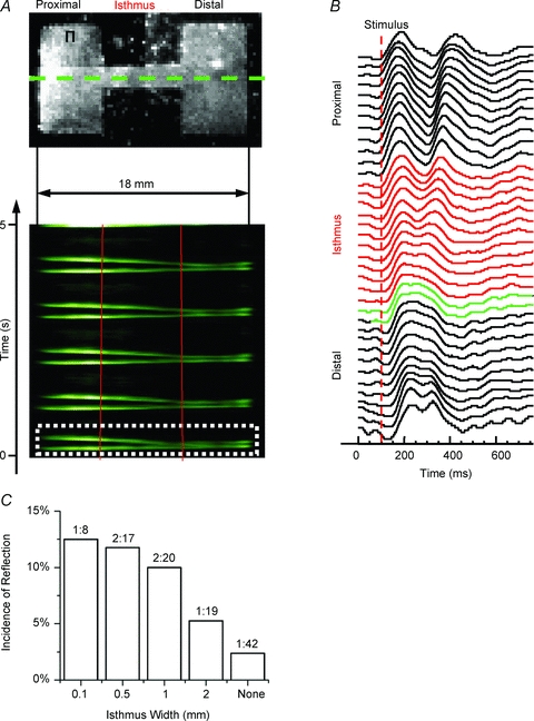
A, top panel, uninfected patterned monolayer (2-mm-wide isthmus) paced at 1 Hz (Π, site of stimulation). A, bottom panel, average TSP for pixels along horizontal lines that traversed the isthmus in A (green dotted line denotes one example). The impulse originates on the upper proximal (left) side and activates the entire preparation distally (right; red lines demark the structural heterogeneity), followed by re-excitation and reflection for each wave. B, optical APs across the preparation for wave 1 (white dotted box in A). Red APs are from pixels within the isthmus and green APs are pixels within the first 500 μm of the distal expansion. C, incidence of reflection at each cable width. Numbers above the bars are the ratio of the number of preparations displaying reflection to the total number of preparations.