
Figure 8. Increased persistent INa promotes EADs and reflection.
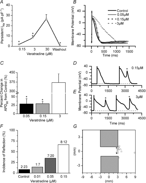
A, change in persistent INa (veratridine minus control) in single NRVMs (*P < 0.05 vs. 30 μm, n = 3–7). B, representative APs at each dosage of veratridine. C, quantification of percentage increase in the APD80 from control (*P < 0.05 vs. 3 μm, n = 3–9). D and E, representative APs and EADs at 0.15 and 3 μm veratridine (dotted line denotes baseline). F, incidence of reflection at each concentration of veratridine (P = 0.003, χ2 test). G, diagram of the monolayer pattern with the site of re-excitation plotted (black square, control; grey circle, 0.01; white triangle, 0.05 μm; and white star, 0.15 μm). One-way ANOVA and Tukey's test were used in A and C.