
Figure 10. Two-dimensional simulations.
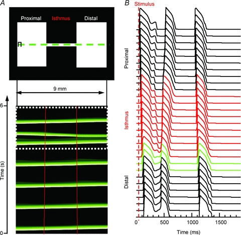
A, top panel, 2D pattern (9 mm × 4.5 mm). A, bottom panel, time–space plot for pixels along the horizontal line that traversed the isthmus in the top panel. The impulse originates on the proximal (left) side and activates the entire preparation distally (right; red lines demark the structural heterogeneitiy). Following the fifth beat, there was re-excitation at the distal expansion region and a retrograde reflected wave. The final beat propagated through the entire preparation without reflection. B, APs across the preparation for the final two beats (white dotted box in A). The red APs are from nodes within the isthmus, and green APs are pixels within the first 600 μm of the distal expansion.