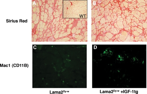
Figure 6.
Fibrosis and inflammation were not noticeably improved by IGF-1 overexpression in Lama2Dy-w muscles. 20× images of Picro-sirius red staining for collagen (red) of (A) Lama2Dy-w and (B) Lama2Dy-w+IGF-1tg TA muscles revealed no noticeable difference in levels of fibrosis. Immunostaining for Mac-1/CD11B (green), a marker of monocytes/macrophages, showed no change in infiltrating inflammatory cells in (C) Lama2Dy-w muscles and (D) Lama2Dy-w+IGF-1tg muscles.

