Figure 3.
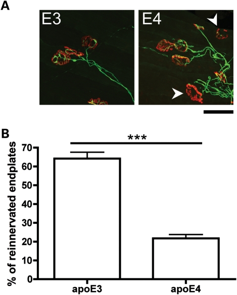
ApoE4 significantly impairs neuromuscular re-innervation following nerve crush injury in the mouse PNS. (A) Representative confocal micrographs showing immunohistochemically labelled NMJs from apoE3 and apoE4 lumbrical muscles 2 weeks after a tibial nerve crush. Pre-synaptic axons and motor nerve terminals are shown in green and post-synaptic acetylcholine receptors labelled with α-BTX are shown in red. Note how the apoE3 endplates have all been re-innervated, whereas some apoE4 endplates are still vacant and awaiting re-innervation (white arrows) at the same time-point. Scale bar = 30 μm. (B) Bar chart (mean ± SEM) showing levels of neuromuscular re-innervation in lumbrical muscles 2 weeks after a tibial nerve crush. Re-innervation was significantly impaired in apoE4 mice compared with apoE3 mice (P< 0.001; Mann–Whitney test, two-tailed; apoE3, n= 5 mice; apoE4, n= 5 mice).

