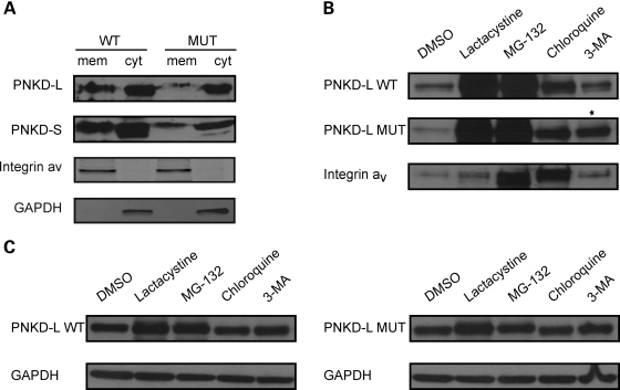
Figure 2.
PNKD-L is degraded through the proteasomal, lysosomal and autophagy pathways. (A) The PNKD protein (upper panel PNKD-L and lower panel PNKD-S) is found in both membrane and cytosolic fractions. (B and C) Cells expressing equal amounts of WT or MUT PNKD-L were treated with DMSO, lactacystine (proteasome inhibitor), MG-132 (proteasome and lysosome inhibitor), chloroquine (lysosome inhibitor) and 3-MA (autophagy inhibitor), respectively, and lysates were extracted into membrane (B) and cytosolic (C) fractions. The membrane-associated WT PNKD-L protein is degraded through the proteasomal and lysosomal pathways (lanes 2–4), PNKD-L MUT is also degraded through the autophagy pathway (lane 5). The star marks the band with increased intensity of PNKD-L MUT proteins versus DMSO control when treated with 3-MA. (C) The cytosolic PNKD-L protein is degraded through the proteasomal pathway (lanes 2 and 3). Integrin αV and GAPDH were used as transmembrane protein and cytosolic protein controls, respectively.

