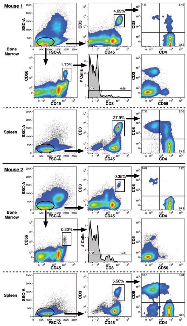
Figure 10.

Detection of human T- and NK-lymphoid cells in mouse bone marrow. T-cells and NK-cells detected in two representative mice are shown. Both lymphoid lineages were defined by expression of low levels of forward- and side-light scatter (oval gates) in addition to gating for PI-CD59+ events excluding mouse cells and doublets as shown in Figure 4. High levels of CD45-FITC expression were also used to help distinguish the CD3–PC7+ T-cells and CD56-PE+ NK-cells (rectangular gates). The respective frequencies of these two lineages among total human CD59+ events are indicated. T-cells were detected in both the BM and among light-density spleen cells. The expression and frequency of CD4–PE and CD8-APC among CD3+CD45+ events are shown. CD56+CD45+ events were only detected in the BM and the frequency of CD8 expression among these events are shown in the histograms. The mutually exclusive expression of CD3 and CD56 in the BM samples is also shown.
