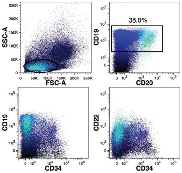
Figure 9.

Detection of human B-lymphoid cells in mouse bone marrow. B-cells were primarily detected using CD19-APC expressing low levels of forward- and side-light scatter (oval gate) in addition to gating for PI–CD59+ events excluding mouse cells and doublets as shown in Figure 4. The percentage of total human CD59+ events expressing CD19 (rectangular region) is indicated. BM cells were also co-stained with CD20-FITC, CD22-PE, and CD34-PC7. Co-expression of these markers is shown using polychromatic dot-plots in which CD19+ events are shown in dark blue and CD19+CD20+ events are shown in light blue, as indicated in the top-right plot. Note the presence of CD19+ events among CD34+ events and the predominance of CD20 expression among CD34– events.
